.gif)


Background and Objective: A cross-sectional study was
conducted to evaluate a clinically relevant position of the
inferior alveolar nerve and canal with respect to the lateral
cortex, alveolar crest, and first and second molar root apices
in adult dentate patients.
Methods: The study was conducted in the patients who
reported various oral and maxillofacial surgical procedures
at the Department of Oral and Maxillofacial Surgery for
whom preoperative computed tomography (CT) scan was
taken for the required surgical procedures. CTs were selected
based on the selection criteria, and a total of 50 CTs were
evaluated. Predictor variables were gender and side. Out-
come variables were the average linear buccal measurements
from the lateral cortex to the outer cortical margin of the
inferior alveolar canal from below the mandibular foramen
till the mental foramen, linear vertical measurements from
the alveolar crest to the superior crest of the inferior alveolar
canal in the third, second and first molar regions, and linear
vertical distance from the superior aspect of inferior alveolar
canal till the apices of second and third molars. Descriptive
statistics were analyzed with t test and paired t test. P value
<0.05 was taken as significant.
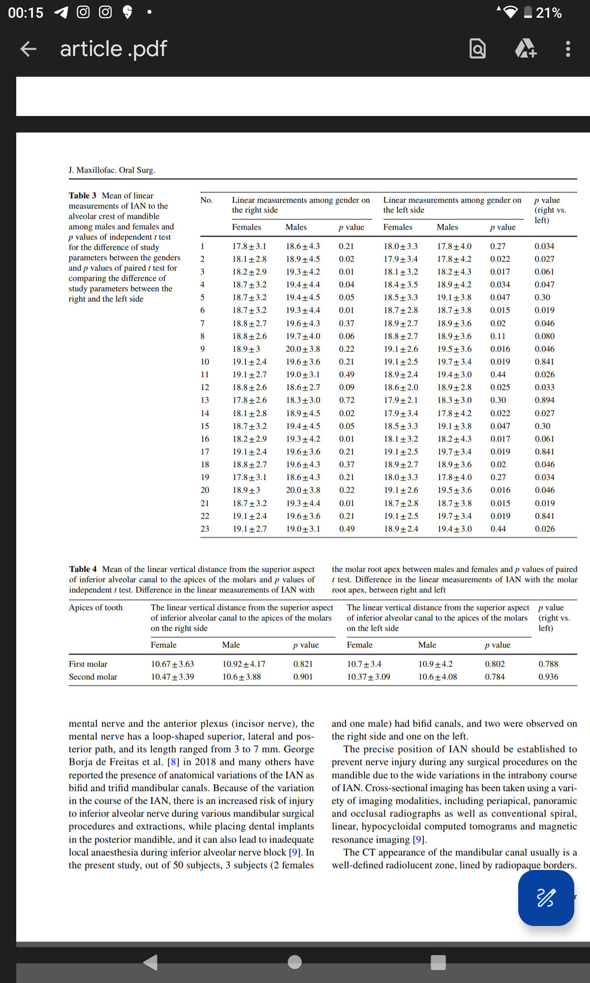
Results: The study sample was composed of 27 male and
23 female patients with a mean age of 29±4.6 years. The
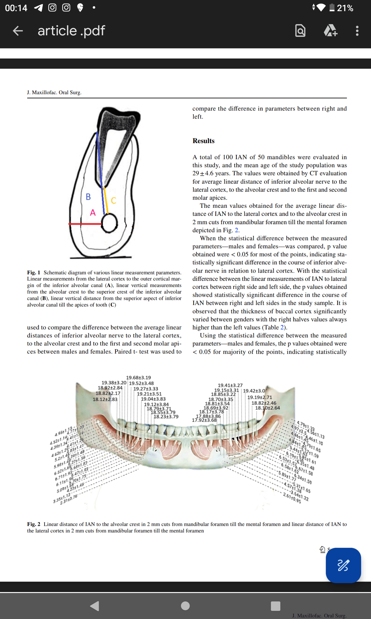
mean value of linear measurements of IAN to the right
lateral cortex at the mandibular foramen was 4.96±1.13,
and for the left side, it was 4.79±1.33. The mean values
of linear measurements from the superior aspect of IAN canal to the alveolar crest at third, second and first molar
regions obtained in this study were 18.5±3.79, 19+3.83
and 19.5+3.48 for the right side and 17.8+3.86, 18.8+3.54
and 19.143.31 for the left side, respectively. The average
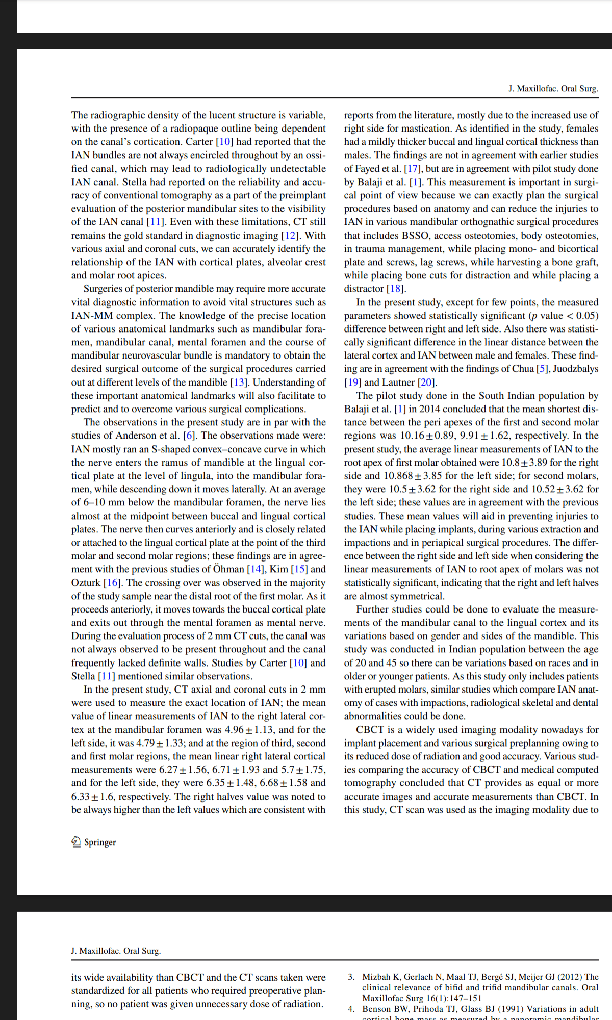
linear measurements of IAN to the root apex of frst molar
were 10.8 3.89 for the right side and 10.868+3.85 for the
left side; for second molars, they were 10.5+3.62 for the
right side and 10.52+3.62 for the left side. Gender and side
infuenced the outcome with varying statistical signifcance.
Conclusion: The study has identified the average linear
measurements of the buccal cortical bone along the path
of IAN in the mandible, average height of alveolar bone
above the IAN and average distance between the IAN with
the first and second molar root apices. This will be useful
guide while planning and performing various mandibular
surgical procedures close to the IAN nerve and procedures
that can disturb the IAN. The implications of these findings
will influence the course of surgery.


Share your thoughts, Get involved!
Comments